Hoof problems
Club foot
Definition club foot
 The club foot is caused by too strong a pull of the deep flexor tendon which pulls the toe wall concave and presses the growth grooves together at the front and pulls them apart at the back. Solution: With a full rocker shoe the pull of the deep flexor tendon is reduced, the concavity of the toe wall disappears and the growth grooves become parallel. A buck hoof is a special form of malpositioning of the horse's limb. In extreme cases, not only the position but also the shape of the hoof is lost. The hoof is too steep to load and use normally.
Types of club foot
In the case of a buck toe, a distinction must be made between a congenital and an acquired buck toe. If a foal is born with a buck hoof, it can usually be corrected completely. In the adult horse, more care must be taken, as the tendons and ligaments are no longer so easily deformable.
Features of the club foot
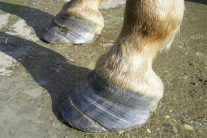
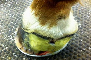
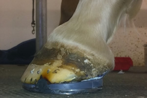
Increased abrasion on the toe, high and steep heels, a horiontal running coronet band, concave dorsal hoof wall, widened white line.
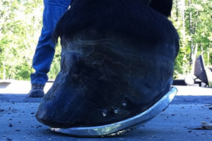
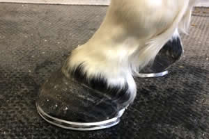
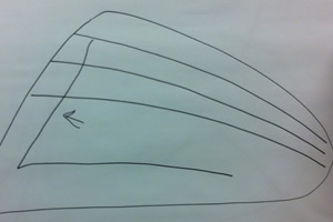
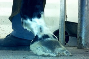
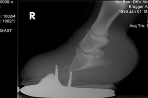
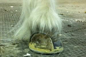
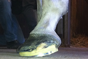
Example club foot, picture 1
Concave toe wall, growth grooves open to the back.
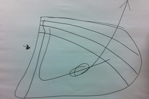
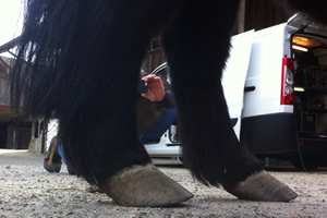
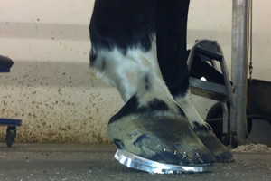
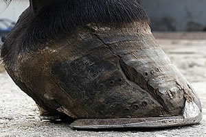
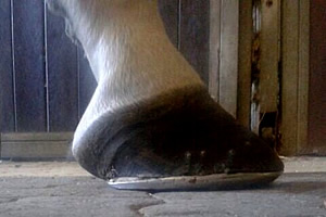
Example club foot, picture 2
Extremely strong club foot
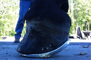
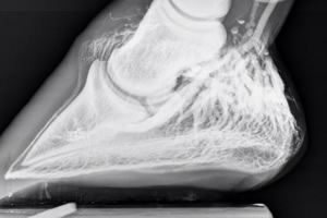
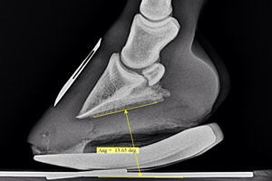
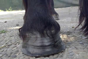
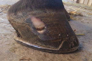
Example club foot, picture 3
Club foot with hoof cleft formation
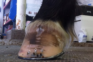
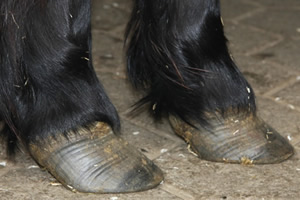
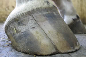
Example club foot, picture 4
Club foot with diseased white line
Example club foot, picture 5
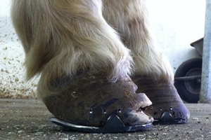
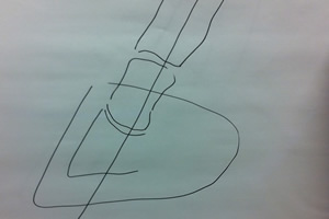
Shoeing variant for club's foot to cancel the pull of the deep flexor tendon
Example club foot, picture 6
Chronic club foot
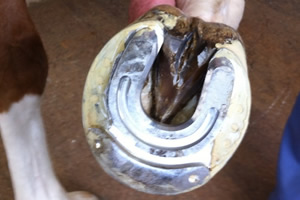
Example club foot, picture 7
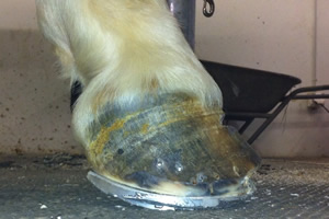
Full rocker shoe for club's foot
Example club foot, picture 8
Hoof brace to widen the heel area
Hoof abscess
Definition hoof abscess
 Accumulation of pus in an all-round enclosed tissue cavity caused by tissue fusion. Hoof abscesses can be very painful and lead to severe lameness.
Cause of hoof abscesses
Extremely cracked and dry hooves, hooves that stand a lot in urine and horse droppings (risk of ammonia decomposition), sharp objects on the pasture or in the exercise area (e.g. broken glass, nails, etc.), improper or infrequent hoof treatment by farrier or hoof groomer, open wounds on the ball or crown, extremely strong impact on sharp stone, too tight horseshoes, incorrect nailing of the iron, thin soft hoof horn, etc.
Treatment of hoof abscess
 With hoof tongs, the place where the pus is located can be felt. With the hoof knife, a funnel-shaped hole is cut at this point so that the pus can drain off. In some cases it is necessary to cut the hole several times over a period of days until the pus has completely drained away. After cutting with the hoof knife, the wound is disinfected.
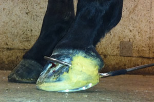
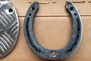
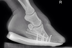
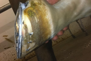
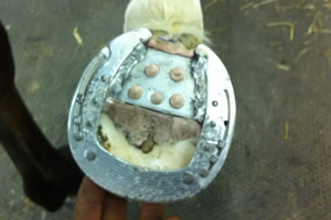
Use of cover irons
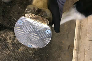
Cover irons are used for large hoof abscesses in the case where the treated horse should be shod again to be led home. This treatment is usually carried out in the veterinary hospital. The advantage of this method is clearly that the wound on the sole side can always be treated again after unscrewing the cover.
Example hoof abscess, picture 1
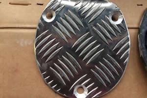
Cover iron
Example hoof abscess, picture 2
Horseshoe for cover iron
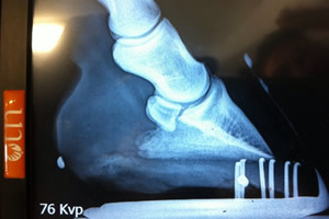
Example hoof abscess, picture 3
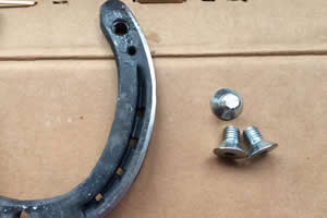
Horseshoe for cover iron with screws
Example hoof abscess, picture 4
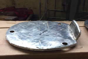
Cover iron
Example hoof abscess, picture 5
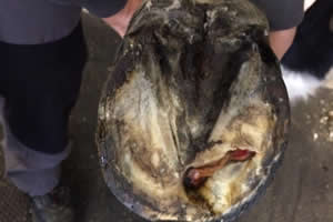
Hoof abscess
Example hoof abscess, picture 6
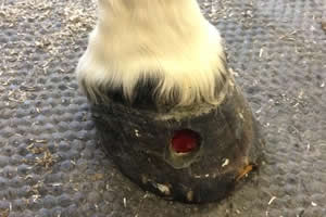
Hoof abscess
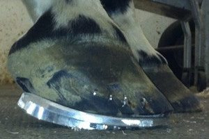
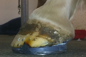
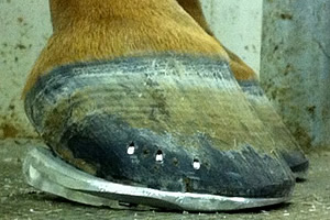
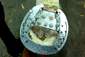
Example hoof abscess, picture 7
Fitting with cover iron
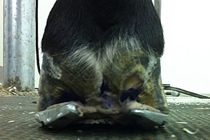
Example hoof abscess, picture 8
Cover iron
Hoof balance
Definition hoof balance
 Ideally, the hoof balance is such that the pressure point - the centre of the hoof joint - falls in the middle of the horseshoe. The same applies, of course, from the front view.
Features hoof balance
When the hoof is optimally balanced, the hoof walls behave straight and symetrically, with no marked change in the growth grooves and no bumps or dents.
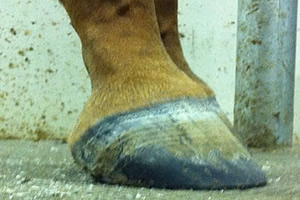
Example hoof balance, picture 1
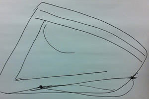
Ideal hoof balance, parallel growth grooves
Example hoof balance, picture 2
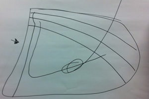
Hoof balance too far forward, growth grooves narrow in front
Example hoof balance, picture 3
Hoof balance too far back, growth grooves narrow at the back
Example hoof balance, picture 4
Ideal hoof balance, straight lateral toe axis
Example hoof balance, picture 5
Extremely flat position, hoof balance too far back
Example hoof balance, picture 6
Ideal hoof balance with balanced blood circulation
Example hoof balance, picture 7
Hoof position too flat
Example hoof balance, picture 8
Correction: By means of Rockerrail fitting
Laminitis
Laminitis
 Once the cause of laminitis has been identified and can be treated, the aim is to relieve the diseased, poorly perfused lamellar zone to prevent rotation of the coffin bone (rocker rail shoeing). This shoe must be applied until the lamellar zone grows down parallel to the tip of the coffin bone, then your horse is healthy again.
Definition laminitis
Laminitis is a disease that occurs in hoofed animals. It is an aseptic diffuse inflammation of the hoof corium, whereby the hoof capsule detaches from the corium. Acute laminitis is an emergency and requires immediate treatment. In extreme cases, it can lead to shoeing. Chronic laminitis can lead to coffin bone rotation.
Types of laminitis
 There are different types of laminitis, such as
- Birth Roe
- Fodder deer
- Stress laminitis
- Poisoning deer
Here is an example of a laminitis treatment I carried out in 2011.
Features of laminitis
 Laminitis can be recognised from a distance and at a glance, so unmistakable are the signs and symptoms of an acute episode of laminitis. The horse walks clammy and stiff as a board, has pain when turning, can walk very badly over hard ground and uneven ground, the facial expression is distorted with pain, the breathing is faster, the pulse faster (from the pain...), sometimes there is even a fever in addition. The symptoms range from mild to moderate to very severe. Not all horses, even with a very massive episode of laminitis, stand in the so-called "roebuck position", this is therefore not an unmistakable sign of laminitis, or rather not a common sign of laminitis, although it is sometimes seen naturally. Laminitis is characterised by excruciating pain, many horses can hardly walk at all, many horses lie down and do not want to get up, every step becomes a torture...
Example laminitis, picture 1
Chronic laminitis with necrotic coffin bone
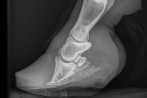
Example laminitis, picture 2
Acute laminitis with severe rotation of the coffin bone
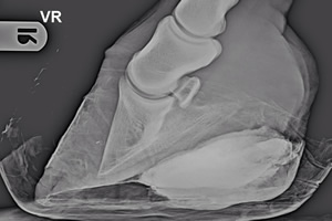
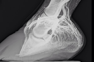
Example laminitis, picture 3
Venogram of chronic laminitis
Example laminitis, picture 4
Acute laminitis with coffin bone rotation
Example laminitis, picture 5
Venogram of chronic laminitis
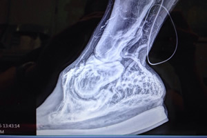
Example laminitis, picture 6
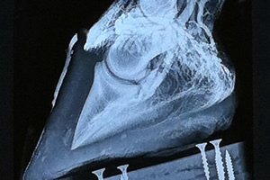
Venogram of acute laminitis
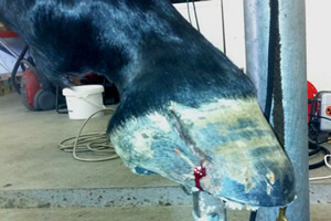
Example laminitis, picture 7
Rockerrail shoeing for acute laminitis
Example laminitis, picture 8
Chronic laminitis
Hoof gap
Hoof gap
 The longitudinal split comes from a crown injury, this is usually not dangerous. A hoof split from the bottom to the top is usually not dangerous either. A hoof split from top to bottom, however, is dangerous. The lateral as well as the hoof balance must be determined exactly from the front. The cause of the tension in the hoof must be identified, then the ideal relieving shoe must be applied and the gap can be glued, screwed or even sewn. A split hoof can lead to lameness if an infection can spread!
Definition hoof gap
Horn splits are cracks in the horse's hoof that run parallel to the horn tubes of the bearing rim. At the beginning of their formation, while they are still small and inconspicuous, they are called wind cracks. They are only called horn cracks when they are so deep that they go all the way through the hoof wall to the hoof corium.
Types of hoof splits
 There are several types of horn splits, namely
- Wind cracks
- bleeding fissure
- Supporting edge gaps
- continuous horn clefts
- Crown margin gaps
It's just an excerpt, of course there are others...
Features hoof splits
 Stumbling, unequal lameness, pain on turning, nothing can be seen externally, only occurs in the front, show jumpers stop in combination.
Example hoof gap, picture 1
Sick white line due to large hoof gap
Example hoof gap, picture 2
Heel hoof gap
Example hoof gap, picture 3
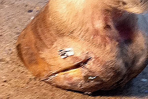
Heel hoof gap
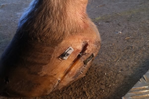
Example hoof gap, picture 4
Hoof split and injury to the hoof corium
Example hoof gap, picture 5

Fixing the heel cleft with a hoof brace
Example hoof gap, picture 6
Bleeding side wall gap, fixed by means of aluminium bridge
Example hoof gap, picture 7
Cross hoof gap due to crown injury
Example hoof gap, picture 8
Fitting variant to take the pressure off the overloaded walls
Position problem
Definition position problem
 In the case of a position problem, we speak of a deviation from the ideal. Ideal means a lateral straight toe axis and a positive palmar angle. The position must be found so that the joint spaces - especially the hoof joint and the coronet joint - behave symetrically.
Types of position problems
There are several types of positional problems, namely
- too steep a position
- Too flat position
- too inclined position
Features Positional problems
 Changes to the growth grooves, namely:
- If the hoof is too steep, the growth grooves are narrow at the front and wide in the heels.
- If the hoof is too flat, the growth grooves are wide at the front and narrow in the heel area.
- in the event of a lateral imbalance, dents and bulges are made in the horn wall and the load grooves are crushed
Example of a position problem, picture 1
Position too flat
Example of a position problem, picture 2
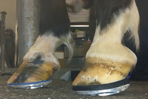
Correction option: Rockerrail fitting
Example of a position problem, picture 3
Toe axle slightly broken forward
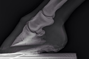
Example of a position problem, picture 4
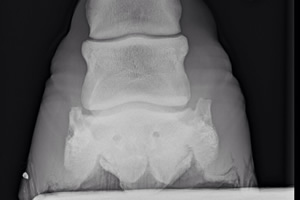
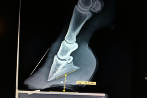
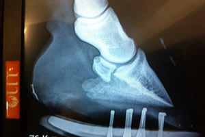
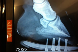
X-ray to assess the joint spaces
Example of a position problem, picture 5
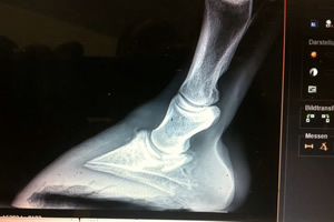
Dramatic negative palmar angle
Example of a position problem, picture 6
Eggplate: possibility for the horse to find its comfortable hoof position
Example of a position problem, picture 7
Hoof length compensation through different thickness underlays
Example of a position problem, picture 8
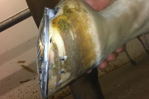
8 degree wedge sole to tip the hoof balance forward
Navicular bone
Navicular bone
 A very good solution for relieving the navicular region is the fullrocker iron (aluminium), with which we increase the palmar angle by up to 8 degrees and relieve the pull of the deep flexor tendon by means of rocker mechanics in relation to the ground.
Definition of the navicular bone
The navicular bone is the sesamoid bone on the deep flexor tendon of the hoof. One navicular bone is formed per toe. It lies under the tendon at the joint between the coffin bone and the coronet bone. Between the tendon and the navicular bone lies a bursa, the hoof roll bursa.
Types of navicular bone
 There are generally four types of fractures affecting the navicular bone, namely
- Chip fractures
- simple fractures
- Rubble fractures
- congenital context separations
Features of the navicular bone
Increased abrasion on the toe, high and steep heels, a horiontal running coronet band, concave dorsal hoof wall, widened white line.
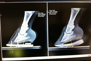
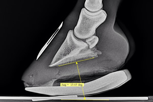
Example navicular bone, picture 1
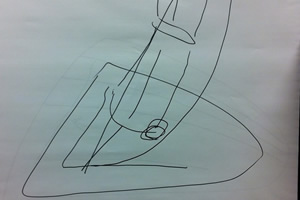
Representation of the navicular bone position when the angle is too flat
Example navicular bone, picture 2
Changing the angle by means of rocker fitting
Example navicular bone, picture 3
Extremely flat fitting without roll-off point, very large jet leg impression
Example navicular bone, picture 4
Correction by means of Rockerrail fitting, extreme relief of the navicular bone
Example navicular bone, picture 5
Too flat fitting without roll-off point, very large navicular pressure
Example navicular bone, picture 6
Too flat fitting without roll-off point, very large navicular pressure
Example navicular bone, picture 7
Fullrocker fitting with light beam pressure
Example navicular bone, picture 8
Fullrocker fitting with light beam pressure
Hoof problems
The following hoof problems are described in more detail here:
FEI HELSINKI 2016
Romain Duguet wins the Longines FEI World Cup in Helsinki with Quorida de Treho - Congratulations. Watch in the video how the iron flies ahead like a bullet...